
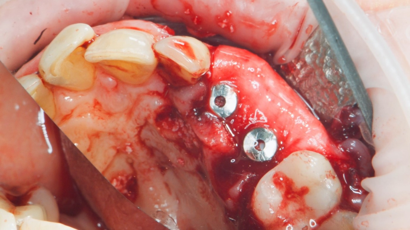

Predictable implant outcomes in patients with systemic conditions that may affect healing depend on sound treatment planning, careful material selection, and a strategy that minimises surgical burden without compromising biological outcomes. Collagen membranes are central to GBR, providing space maintenance and barrier function throughout the healing phase. In challenging clinical situations—multi-tooth defects, thin buccal walls, or cases requiring membrane fixation under tension—membrane stability and handling characteristics become critical factors.
Geistlich Bio-Gide® Forte (Geistlich Pharma) is a resorbable collagen membrane with high tensile strength and enhanced form stability upon hydration, building on the established biological performance of Geistlich Bio-Gide®. In cases involving medically compromised or elderly patients, where healing capacity may be reduced, additional biological support can be beneficial. REGENFAST® (Mastelli) is a viscoelastic gel containing polynucleotides and hyaluronic acid and is designed to support cellular processes involved in tissue repair and wound healing. This case report describes the combined use of both materials in a clinically demanding scenario.
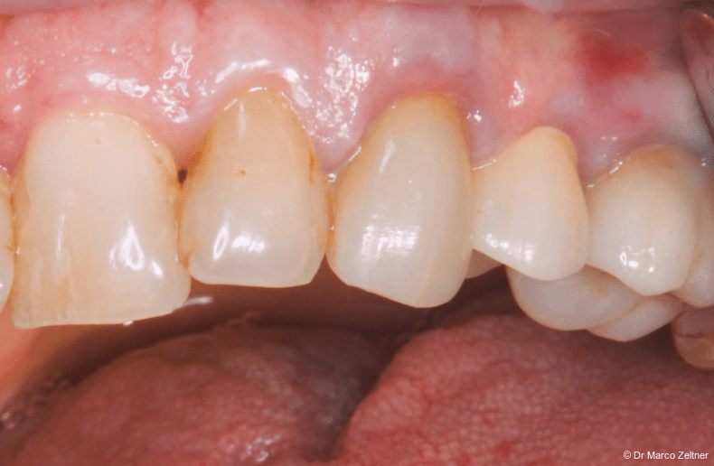
Fig. 1a: An elderly patient presented with loose crowns and apical infections affecting teeth #23 and #24. Both teeth had a hopeless prognosis, and the crowns were removed without resistance.
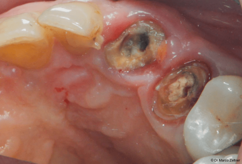
Fig. 1b: An elderly patient presented with loose crowns and apical infections affecting teeth #23 and #24. Both teeth had a hopeless prognosis, and the crowns were removed without resistance.
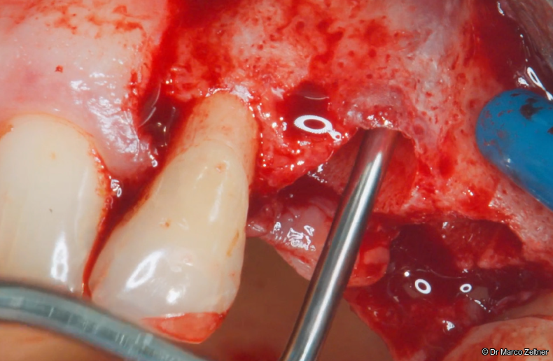
Fig. 2: Clinical examination revealed an extremely thin buccal bone wall.
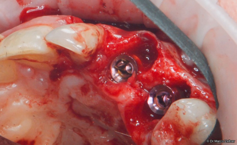
Fig. 3: Tissue-level implants measuring 4.1 × 10 mm (Matrix®, TRI) were placed immediately at sites #23 and #24 using a guided surgical approach.
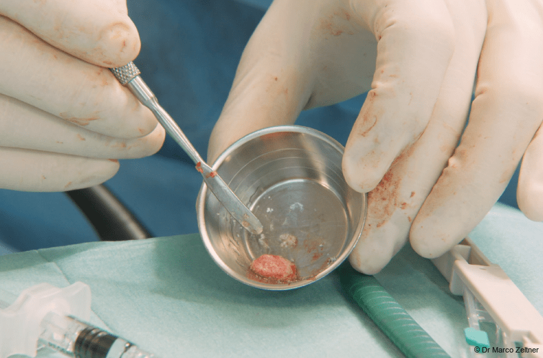
Fig. 4: Autologous bone was combined with 0.5 g of small Geistlich Bio-Oss® granules and REGENFAST® to promote faster healing.
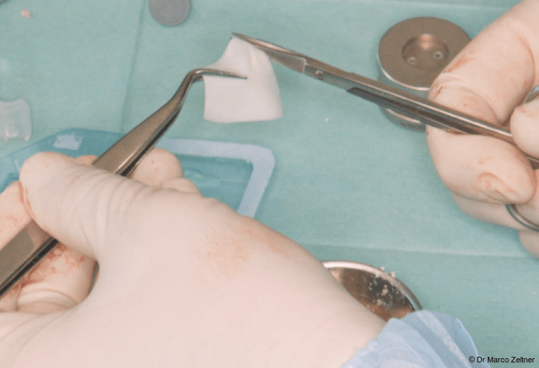
Fig. 5: The Geistlich Bio-Gide® Forte membrane was hydrated with sterile saline and trimmed to conform to the defect morphology.
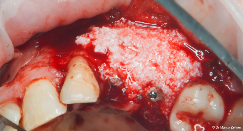
Fig. 6: The defect was grafted with the mixture of autologous bone, Geistlich Bio-Oss®, and REGENFAST®.
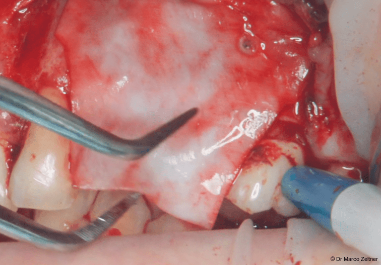
Fig. 7a: The membrane was positioned over the graft and stabilised with resorbable pins.
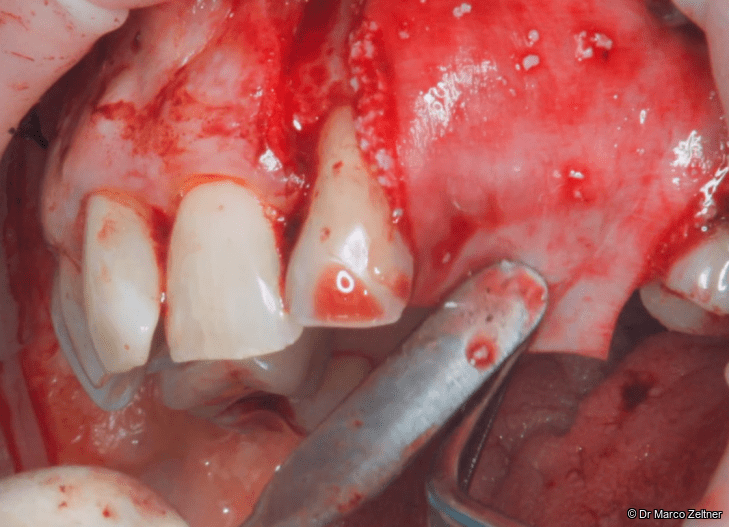
Fig. 7b: The membrane was positioned over the graft and stabilised with resorbable pins.
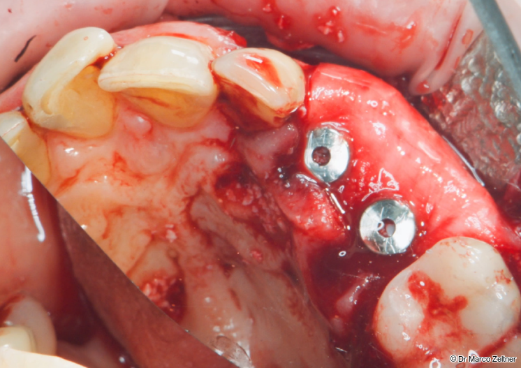
Fig. 8: A small incision was made in the membrane to adapt it around the healing abutments. REGENFAST® was then applied to the Geistlich Bio-Gide® Forte membrane.
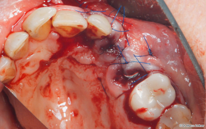
Fig. 9a: The surgical site was closed using two horizontal mattress sutures and additional single interrupted sutures.
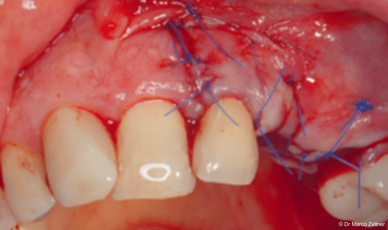
Fig. 9b: The surgical site was closed using two horizontal mattress sutures and additional single interrupted sutures.
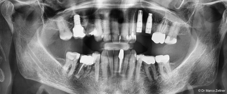
Fig. 10: Postoperative radiographic assessment.
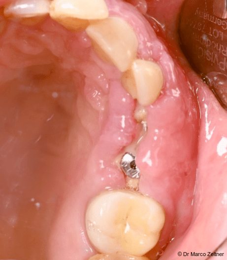
Fig.11a: Soft-tissue healing following surgery.
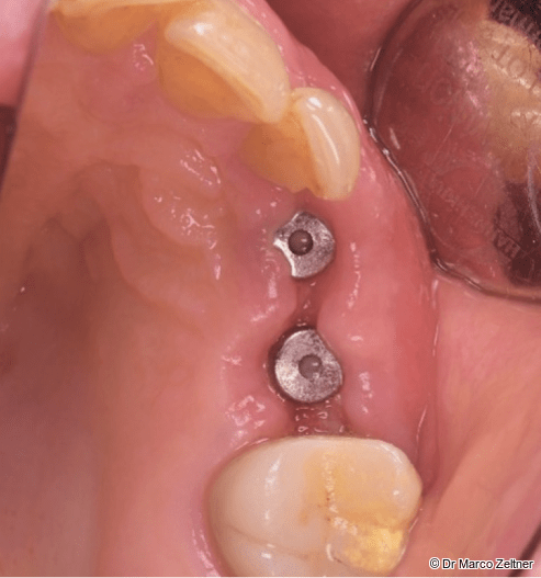
Fig. 11b: Soft-tissue healing following surgery.
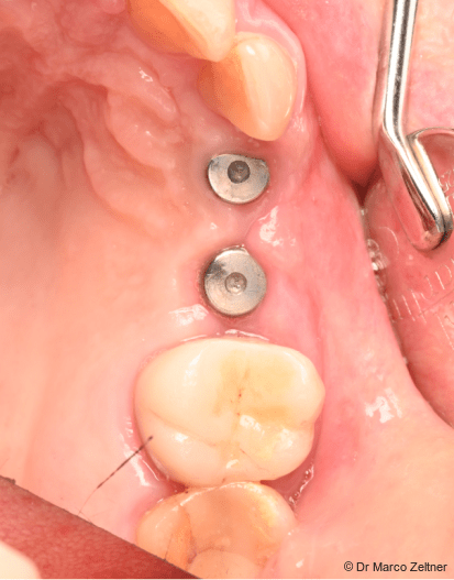
Fig. 12: Healed soft tissues and successful implant osseointegration before intraoral scanning for fabrication of the definitive prosthetic restoration.
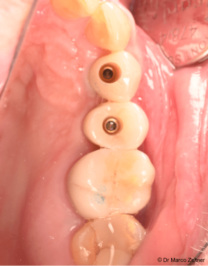
Fig. 13: Five months post- operatively, with the final prosthetic restoration in place.
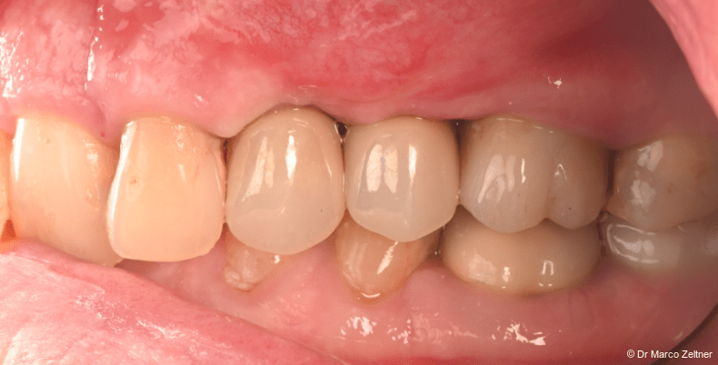
Fig. 14: Lateral view of the final prosthetic restoration in place, demonstrating the resulting occlusal relationship.
Case report
Initial situation
A 78-year-old female nonsmoker, with no history of hospitalisation within the preceding six months, presented with pain, mobile crowns, and clinical signs of an apical infection involving teeth #23 and #24. CBCT revealed a subgingival coronal fracture of tooth #23 and a subgingival coronal fracture associated with apical osteitis at tooth #24. Following comprehensive clinical and radiographic evaluation, both teeth were
considered non-restorable and assigned a hopeless prognosis. The crowns were removed without resistance (Figs. 1a+b), followed by extraction of the affected teeth. Subsequent clinical inspection of the extraction sites revealed an extremely thin buccal cortical plate (Fig. 2).
The patient’s medical history included anticoagulation therapy and antihypertensive medication. Given her systemic status and preference for limiting surgical visits, treatment planning prioritised efficiency while maintaining regenerative quality.
Treatment planning
Immediate implant placement was planned based on the absence of acute suppurative infection, the availability of sufficient residual
bone to achieve primary implant stability, and the patient’s medical profile, which favoured minimising the number of surgical interventions. Both teeth were therefore atraumatically extracted, and the implants were placed during the same surgical session, obviating the need for a staged approach.
The simultaneous extraction of two adjacent teeth was expected to result in a non-self-stabilising osseous defect, where defect morphology poses a challenge and graft stability depends on a stabilising element. A slowly resorbing bone substitute, autogenous bone, a biologic adjunct, and a high-strength collagen membrane were selected to achieve reliable augmentation and support healing in this medically compromised patient.
Surgical procedure
Following administration of local anaesthesia, teeth #23 and #24 were extracted using an atraumatic technique. A full-thickness mucoperiosteal flap was elevated through a horizontal crestal incision and a single vertical releasing incision mesial to tooth #22. The extraction sockets were then thoroughly debrided. Immediate implant placement was performed at sites #23 and #24 (Fig. 3) using 4.1×10mm tissue-level implants (Matrix® implants with a P45 platform, TRI). Both implants were placed using guided surgery and inserted with 25 Ncm torque. This value was sufficient for transmucosal healing but did not support immediate loading; healing abutments were placed on both implants to avoid further surgical access.
The osseous defect was grafted with a mixture of autogenous bone (harvested with a Safescraper® (META Technologies), 0.5 g of Geistlich Bio-Oss® small granules (Geistlich Pharma), and REGENFAST® to support bone regeneration and soft-tissue healing (Figs. 4–6).
Geistlich Bio-Gide® Forte was hydrated with sterile saline and trimmed to the defect (Figs. 7a+b). Two small cuts were made to position the membrane around the healing abutments (Fig. 8). The membrane was stabilised with LeadFIX resorbable pins and the healing abutments, ensuring stable coverage of the augmented volume. REGENFAST® was additionally applied on top of the membrane (Fig. 8). The flap was closed with SERALON® 4 and 5 non-resorbable sutures using two horizontal mattress and single interrupted sutures (Figs. 9a+b).
Postoperative medication consisted of amoxicillin 750 mg three times daily for five days, Dafalgan® as needed for analgesia, and a 0.12% chlorhexidine mouthrinse supplemented with hyaluronic acid.
Postoperative course and follow-up
Postoperative radiographic imaging confirmed satisfactory implant position and osseous fill (Fig. 10). The postoperative course was uneventful with very good soft tissue healing—a notable result given the patient’s age and medical status (Figs. 11–12). Prosthetic restoration followed five months after surgery. The bone and soft-tissue conditions, including the emergence profile, were highly favorable, allowing for straightforward placement of the crowns.
Discussion
This case illustrates a clinical scenario where the goal of reducing surgical burden shaped every decision, from treatment planning to material selection. A 78-year-old patient receiving anticoagulant and antihypertensive therapy and presenting with two adjacent, non-restorable teeth would not generally be considered an ideal candidate for immediate implant placement.
Yet the alternative—staged extraction and delayed implant placement—would have required additional surgical appointments, additional anaesthesia, and a longer overall treatment timeline. Given her systemic profile and her own preference, this was not the preferred path.
The key regenerative challenge was defect morphology. The simultaneous extraction of two adjacent teeth results in a non-contained defect, where graft stability cannot be achieved without a reliable stabilising element. In this case, the membrane needed to be pinned and simultaneously adapted around the healing abutments, requiring sufficient mechanical stability to minimise tearing or deformation. Geistlich Bio-Gide® Forte could be securely pinned and shaped around the abutments without tearing and maintained its form upon hydration, facilitating stable positioning.
The choice to support regeneration with autogenous bone, Geistlich Bio-Oss®, and REGENFAST® was driven by both the biological demands of the defect and the patient’s age. In older patients, healing conditions are less favourable and regenerative potential is reduced. REGENFAST® was incorporated into the graft and applied over the membrane as a biological adjunct, with the aim of supporting cellular processes involved in tissue repair and wound healing*, which may be particularly relevant in patients with reduced regenerative capacity.
The predictable handling of Geistlich Bio-Gide® Forte—stable, trimmable, and tear-resistant—allowed the procedure to be completed efficiently. In a patient where reducing surgical time and complexity matters, this enabled us to provide a very patient-friendly approach.
Conclusion
This case demonstrates that immediate implant placement combined with simultaneous GBR can be clinically viable even in older, medically compromised patients, provided that regenerative materials are selected to meet the specific biological and biomechanical demands of the clinical situation. Geistlich Bio-Gide® Forte provided the membrane stability required to manage a non-self-stabilising defect; REGENFAST® provided additional biological support in a clinically challenging healing environment. The outcome demonstrates that a well-planned approach can reduce treatment burden without sacrificing regenerative predictability.
* Beretta M, et al.: Int J Periodontics Restorative Dent 2025 Mar 6;45(2): 209–219.